Hiatal Hernia
Hiatal hernias are common in French Bulldogs and other brachycephalic (short-nosed) breeds. It is a complex disease involving a congenital malformation of the diaphragm which results in herniation of the stomach into the thorax and chronic regurgitation. It is very commonly associated with brachycephalic airway syndrome which is another set of congenital abnormalities which results in narrow upper airways and breathing difficulties.
Anatomy
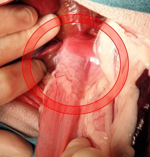
Intra-op image of oesophageal hiatus in a French Bulldog with deficient diaphragmatic muscle around the hiatus
The oesophageal hiatus is the hole in the diaphragm where the oesophagus passes from the thorax into the abdomen to insert into the stomach. It is normally surrounded by the muscle of the diaphragm which forms a physical barrier to prevent the stomach entering the thoracic cavity (chest). The most common form of hiatal hernia (type 1) involves the stomach sliding back into the oesophagus because of an enlarged hiatus which is a congenital malformation (present at birth). The less common form (type 2) is a paraoesophageal hernia where the stomach slides next to the oesophagus into the thoracic cavity.
Signs of hiatal hernia
Most dogs with hiatal hernia will have a history of intermittent or consistent regurgitation after eating. Regurgitation is a passive process where the food literally bounces back up the oesophagus. This differs from vomiting which is an active process involving contraction of the stomach. Mildly affected dogs may initially experience subclinical regurgitation which results in repeated damage to the oesophagus by stomach acid and oesophagitis (heartburn). Eventually this will lead to oesophgeal dysfunction and regurgitation. More severely affected dogs may regurgitate so often that they eventually breathe in (aspirate) their stomach contents and develop aspiration pneumonia which can be life threatening.
How is a hiatal hernia diagnosed?
Often the diagnosis can be made based on breed and history alone (ie. French Bulldog with a history of regurgitation) and by ruling out other systemic causes via blood tests and abdominal ultrasound. In other cases, or in those refractory to surgical management, the patient may require radiographs or endoscopic assessment of the stomach and upper small intestine to rule out other causes of chronic gastrointestinal disease.
Brachycephalic airway syndrome is often diagnosed concurrently and may contribute to the degree of herniation as the dog creates a large negative pressure in its chest to breathe drawing air into its lungs but also drawing its stomach into its chest.
How is a hiatal hernia treated?

Intra-op image of oesophageal hiatus in a French Bulldog after phenoplasty and oesophagopexy
Surgical management of hiatal hernia involves evaluation by direct surgical exposure of the oesophageal hiatus. The most common treatment involves:
Phenoplasty - plication (tightening) of the oesophageal hiatus with non-absorbable suture
Oesophagopexy – suturing the abdominal oesophagus to the diaphragm to prevent it sliding into the thorax
Post-operative care
Most patients will be discharged within one to two days of surgery and there is usually a rapid improvement in the frequency of regurgitation, often with complete resolution of clinical signs. Some dogs may require ongoing medical management, especially if they have other causes of GI disease such as inflammatory bowel disease or food allergies.